

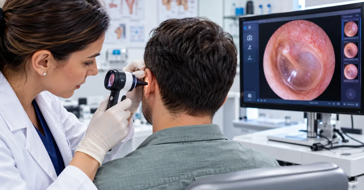
An ear examination is a clinical diagnostic procedure that inspects the outer ear, auditory canal, and tympanic membrane using an illuminated instrument called an otoscope. Doctors, audiologists, and ear specialists evaluate structural tissue integrity, clear obstructive cerumen impaction, identify fluid accumulation behind the eardrum, and detect middle ear infections. Modern digital otoscopes improve this process by transmitting high-magnification video to digital displays, allowing physicians to record time-stamped images for patient tracking and specialist referrals.
What Happens During a Professional Ear Examination
During a routine ear exam, a clinician evaluates the outer ear pinna, inspects the auditory canal for obstructions, and examines the eardrum using an illuminated magnifying diagnostic otoscope. The doctor assesses eardrum color, translucency, vascularization, and structural landmarks like the malleus bone to detect middle ear fluid, pressure imbalances, or acute infections.
- The clinician reviews symptoms and checks the external pinna, postauricular tissue, and tragus for signs of localized swelling, redness, or discharge. Tenderness upon pressing the tragus usually points to otitis externa, while mastoid tenderness flags potential deeper complications.
- The examiner straightens the natural curve of the ear canal by gently pulling the adult pinna upward and backward. For young infants and toddlers, the pinna is retracted downward and backward to align with their shorter, differently angled anatomy.
- The provider selects the largest comfortable disposable speculum tip to maximize lighting and field of view, then gently guides the instrument into the outer cartilaginous third of the canal without touching the sensitive bony canal wall.
- The physician performs a thorough eardrum inspection, looking for a healthy pearly-gray coloration, an intact triangular cone of light reflection in the anterior inferior quadrant, and the visible handle of the malleus. Any amber fluid line, redness, or outward bulging indicates active pathology.
- The clinician attaches a rubber insufflation bulb to perform pneumatic otoscopy whenever fluid entrapment is suspected. A healthy membrane flexes smoothly against controlled air pulses, whereas an eardrum weighed down by middle ear effusion remains rigid.
How Digital Otoscopes Compare to Traditional Handheld Otoscopes
A digital otoscope differs from a traditional handheld otoscope by replacing tiny monocular viewing lenses with digital camera sensors and high-definition video displays. While conventional otoscopes limit visual inspection to the doctor’s immediate memory, an otoscope with camera streams live magnified video, captures permanent visual records, and supports side-by-side historical comparisons across patient visits.
The primary difference between the two systems centers on visual documentation and clinical recall. Standard optical instruments force the clinician to squint through a small viewport and write subjective notes into a paper or electronic file. Once the scope leaves the ear canal, no objective visual record remains. A modern digital ear otoscope solves this limitation by archiving high-resolution image files or video clips directly into electronic health record systems. This creates a verifiable audit trail that protects the provider legally and allows direct visual comparisons across subsequent visits.
Patient engagement and clinical magnification also set these technologies apart. Standard optical tools isolate the view so that only the examining clinician can observe the eardrum. An otoscope with screen projects the examination live onto an external tablet or clinic monitor, enabling the doctor to show patients or parents the exact cause of discomfort, whether it is an inflamed canal or a foreign object. High-end hd video otoscope hardware also provides up to 30x digital magnification, making fine structural tears and subtle vascular changes far easier to spot than through traditional pocket instruments.
What Medical Conditions Does an Otoscopic Ear Checkup Detect
An otoscopic ear checkup detects acute otitis media, outer ear canal infections, ear wax impaction, eardrum perforations, foreign body blockages, and cholesteatomas. By inspecting the tympanic membrane and ear canal skin, physicians differentiate mechanical sound obstructions from nerve-related auditory damage that requires formal audiologist hearing tests.
Cerumen impactions represent the most frequent finding during routine checkups. When earwax solidifies into an occlusive plug, it directly causes conductive hearing loss, physical fullness, and tinnitus. Otoscopy confirms whether manual extraction, water irrigation, or microsuction is necessary to safely clear the canal.
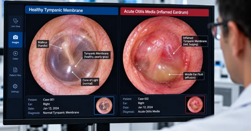
Beyond simple wax blockages, an otoscope reveals deep middle ear pathologies. In cases of acute otitis media, the eardrum appears fiery red, opaque, and visibly bulging outward from trapped purulent fluid. Serous otitis media presents differently, showing an amber discoloration with distinct air bubbles trapped behind a retracted membrane. The examination also detects dangerous chronic conditions such as tympanic membrane perforation following acoustic trauma, or destructive cholesteatoma cysts that form in the upper attic region of the eardrum and risk eroding the delicate bones of hearing.
How Pediatric Ear Examination Differs for Infants and Children
A pediatric ear examination requires smaller speculum sizes, specialized physical positioning, and modified canal-straightening techniques due to developing anatomical differences. Infants possess shorter, more horizontal ear canals and Eustachian tubes, making young children significantly more prone to rapid fluid accumulation, painful middle ear infections, and recurrent otitis media.
Young children rarely sit still during an ear exam, which creates an immediate risk of canal trauma if the patient jerks their head. Clinicians overcome this by having the parent position the child securely on their lap, bracing the child’s head firmly against the parent’s chest while holding both arms. The physician then anchors their own hand against the child’s cheek while holding the otoscope like a pen, ensuring that any sudden head movement carries the instrument safely with the head rather than jabbing into the canal wall.
Anatomical differences also require an inverted traction technique. While an adult ear canal curves in an S-shape that straightens with an upward and backward pull, an infant canal runs in the opposite direction and straightens only when the pinna is drawn downward and backward. Using a digital otoscope for children transforms these visits because modern camera sensors capture a sharp, frozen frame in a fraction of a second. This allows the doctor to inspect a restless child’s eardrum without prolonged restraint, while showing parents why antibiotics or tympanostomy tubes are clinically necessary.
What Does an Ear Examination Cost and What Are Equipment Price Tiers
A routine clinic ear examination cost ranges between ₹300 to ₹1,500 INR ($50 to $150 USD) depending on whether wax removal or audiometric screening is required. For healthcare facilities purchasing diagnostic equipment, the digital otoscope price ranges from ₹3,000 to ₹25,000 INR ($40 to $300 USD) for entry-level USB and mobile scopes, and up to ₹35,000 to ₹1,80,000+ INR ($500 to $2,500+ USD) for a professional digital otoscope.
Healthcare facilities and private practitioners choose equipment across three distinct investment levels based on their patient volume and imaging requirements.
Entry-level USB and mobile otoscopes cost between ₹3,000 and ₹15,000 INR ($40 to $200 USD). These compact tools feature miniature camera sensors and integrated LED light rings that connect directly to smartphones, tablets, or laptops over USB or local Wi-Fi. They serve well for remote home visits, rural field triage, and simple visual checks by visiting home care nurses.
Mid-range diagnostic instruments manufactured by medical brands like Welch Allyn and Heine range from ₹25,000 to ₹75,000 INR ($350 to $1,000 USD). These clinical models incorporate optical-grade glass lenses, high-output fiber-optic illumination, swappable pediatric tips, and dedicated software drivers that integrate directly with clinic management software.
Specialized ENT diagnostic towers represent the top tier, costing between ₹1,00,000 and ₹3,50,000+ INR ($1,500 to $4,500+ USD). Designed for high-volume outpatient centers and surgical clinics, these video suites combine high-definition video feeds, pneumatic insufflation controls, integrated suction systems, and surgical microscopy tools for planning complex reconstructive middle ear procedures.
How Telehealth Workflows Utilize Digital Otoscopy
Modern telehealth otoscope workflows enable general healthcare workers to capture high-definition images or videos of the eardrum and securely transmit them to remote ENT specialists. This store-and-forward teletriage model reduces unnecessary patient travel, speeds up specialist diagnostic reviews from weeks to hours, and delivers accurate ear evaluations to rural or underserved communities.
Patients in rural or community clinics often wait weeks to secure an in-person consultation with an ear specialist. During that waiting period, simple middle ear effusions can harden or progress into perforated chronic infections.
Equipping general practitioners with digital diagnostic instruments eliminates this bottleneck. A primary care nurse positions the device inside the ear canal, records a five-second video sweep of the tympanic membrane under pneumatic pressure, and attaches the clip to the patient’s encrypted electronic file.
The remote otolaryngologist opens the file on a high-resolution clinic workstation, examines the structural integrity of the pars tensa and attic retraction pockets, and provides an immediate treatment directive. This triage model keeps straightforward cases managed locally within primary care facilities while prioritizing genuine surgical emergencies for direct hospital admission.