

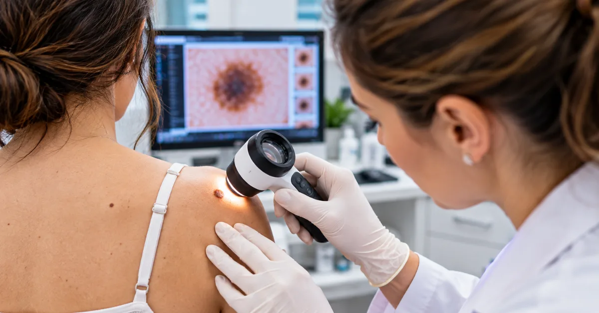
Digital dermoscopy is an advanced non-invasive skin examination method that uses digital dermascopes to magnify, illuminate, and capture high-resolution imagery of skin lesions for baseline documentation, long-term monitoring, and automated diagnostic support. By combining cross-polarized lighting with electronic archiving, digital systems reveal subsurface pigment networks that remain invisible during traditional naked-eye exams. This approach helps clinicians identify early-stage melanomas, track structural mole changes, reduce unnecessary surgical biopsies, and streamline referral workflows across modern healthcare practices.
What Is Digital Dermoscopy and How Does It Work
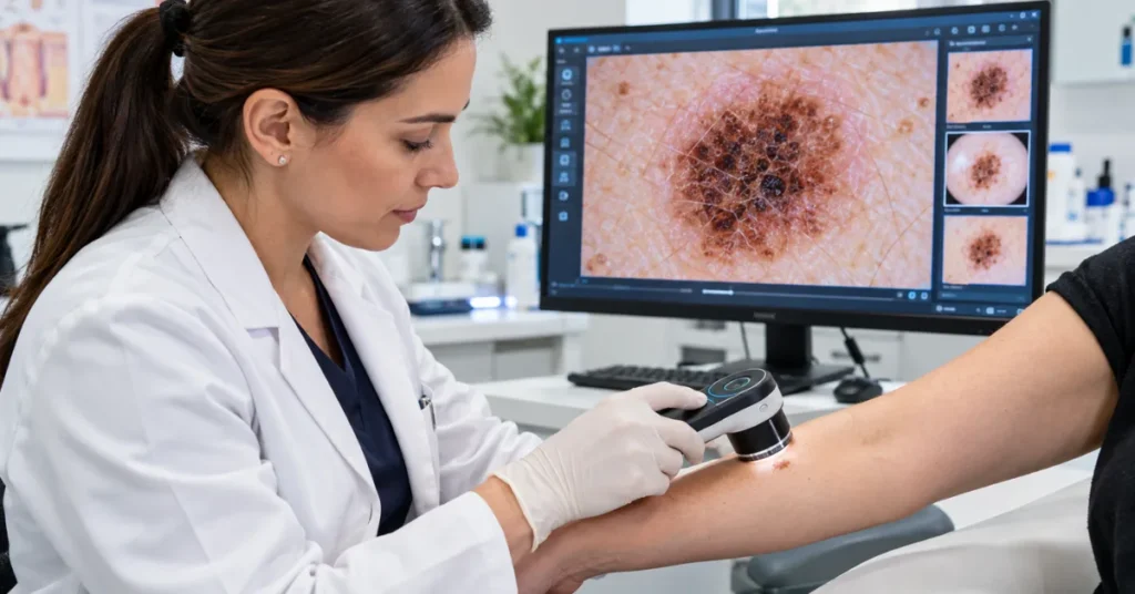
Digital dermoscopy works by capturing magnified skin imagery through specialized optical lenses and cross-polarized light-emitting diodes that eliminate surface glare from the stratum corneum. The digital dermatoscope transmits high-resolution images of subsurface epidermal and dermal structures directly to connected clinical software, where doctors timestamp, measure, and analyze pigmentation patterns against existing patient records.
Traditional visual screening depends entirely on what a clinician sees under an ambient examination lamp. Digital systems eliminate visual guesswork through four primary technical stages.
- Optical Magnification and Glare Reduction
The device uses multi-element glass lenses providing 10x to 40x optical magnification. Polarized light arrays cancel out epidermal surface reflections, allowing clear visualization of deeper structures such as melanin pigment networks, vascular patterns, and crystalline streaks without requiring liquid immersion oils. - High-Resolution Digital Image Capture
An integrated digital sensor records macro-level photographs of targeted lesions. These raw visual files capture border geometry, asymmetry, and color distribution at sub-millimeter clarity. - Archival and Electronic Record Storage
Captured files integrate directly into Electronic Medical Record systems or dedicated dermatology imaging platforms. Each image file contains standardized metadata including capture date, exact anatomical location, and clinical notes. - Algorithmic and Pattern Flagging
Modern digital dermoscopy platforms run analytical models that scan images for irregular structural criteria. The software flags suspicious architectural patterns, helping clinicians decide whether a lesion requires immediate excision or ongoing clinical observation.
What Is the Difference Between a Dermatoscope and a Dermascope
The difference between a dermatoscope and a dermascope comes down to formal medical taxonomy versus popular commercial and aesthetic phrasing. A dermatoscope, also known as a dermoscope, is a regulated medical diagnostic device designed for dermatologists to identify skin cancers. In contrast, dermascope is a common trade spelling often used by aesthetic clinics, beauty spas, or low-cost USB microscope manufacturers.
Physicians and oncologists use the terms dermatoscope, dermoscope, and epiluminescence microscope interchangeably when referring to certified medical diagnostic tools.
Search queries often blend these terms, leading to confusion when clinics shop for clinical equipment. High-grade clinical devices feature verified optical glass, calibrated color rendering indexes, and certified electromagnetic shielding. Basic aesthetic dermascopes often use plastic lenses and uncalibrated white LEDs that distort subtle pigment patterns, making them unsafe for actual skin cancer screening.
How Does Digital Dermoscopy Compare to Traditional Handheld Dermoscopy
Digital dermoscopy differs from traditional handheld dermoscopy primarily through permanent digital record keeping and side-by-side comparative analysis. Traditional dermoscopy relies entirely on a doctor’s immediate memory of what a mole looked like during a past visit, whereas digital dermoscopy creates a permanent, time-stamped visual history of every lesion.
Choosing between traditional analog tools and digital imaging platforms involves clear clinical trade-offs across four operational areas.
- Long-Term Lesion Monitoring
Traditional tools offer zero image recall once the lens lifts off the patient. Digital dermascopes store baseline images, allowing clinicians to overlay past and current photographs to spot tiny micro-architectural changes over time. - Documentation and Legal Protection
Traditional examinations rely on subjective written notes or rough hand-drawn body maps in physical charts. Digital devices build an objective, time-stamped visual audit trail that provides clear clinical documentation if questions arise later. - Diagnostic Speed and Portability
Handheld analog units fit directly inside a lab coat pocket and operate instantly without boots, cords, or software logins. Digital platforms require computer interfaces, wireless pairing, or camera docks, adding slight handling overhead to routine patient checks. - Patient Trust and Consultation Quality
Traditional dermoscopy leaves patients in the dark while the clinician looks through an eyepiece. Digital units display the magnified lesion live on a tablet or desktop monitor, allowing the clinician to educate the patient and justify biopsy decisions visually.
How Does Sequential Digital Dermoscopy Imaging Detect Melanoma Early
Sequential digital dermoscopy imaging detects early melanoma by identifying subtle architectural changes in suspicious moles over structured 3-month and 6-month intervals. This clinical protocol tracks dynamic changes in border shape, asymmetric pigment distribution, and size that indicate malignancy in early-stage melanomas that otherwise appear harmless on day one.
Many aggressive or early-stage melanomas lack the classic asymmetry and color irregularities seen in advanced skin cancers. To solve this clinical challenge, dermatologists use Sequential Digital Dermoscopy Imaging, or SDDI, to evaluate biological activity over time.
Short-term sequential monitoring focuses on single atypical lesions that display ambiguous dermoscopic features. Clinicians schedule follow-up appointments at three-month intervals. Clinical evidence demonstrates that over ninety-five percent of changing melanomas demonstrate visible structural shifts within this 90-day window. If an atypical mole changes in pigment layout, it is flagged for immediate surgical excision. If it remains unchanged, it is safely classified as benign and spared from unnecessary surgery.
Long-term surveillance and mole mapping serve high-risk individuals, particularly patients with dysplastic nevus syndrome or a family history of melanoma. These patients undergo total body photography paired with digital dermoscopy every six to twelve months. Calibrated software matches every mole against baseline photographs, instantly highlighting newly formed lesions or enlarging borders across large surface areas.
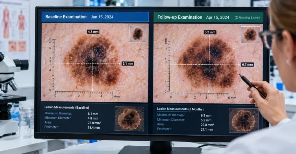
This sequential imaging workflow protects patients in two directions. It catches featureless melanomas before they invade deeper tissue layers, while simultaneously preventing healthy patients from undergoing painful, scarring biopsies on harmless stable moles.
Can a Digital Dermatoscope Detect Skin Cancer Without a Biopsy
A digital dermatoscope cannot confirm skin cancer without a biopsy because non-invasive surface optics cannot analyze cellular pathology beneath tissue layers. While a digital dermatoscope significantly increases diagnostic accuracy and identifies suspicious patterns, surgical excision and laboratory histopathology remain the only definitive legal and medical confirmation of malignancy.
Patients frequently ask if a digital scan or software algorithm can replace a physical scalpel. The answer is firmly no, and the clinical workflow explains why.
A digital dermascope acts as an optical risk assessment filter. It tells a trained physician whether a mole carries high or low statistical odds of being cancerous. When a lesion exhibits suspicious dermoscopic features such as atypical pigment networks, blue-white veils, or irregular peripheral dots, the clinician marks it for surgical removal.
The excised tissue is sent directly to a pathology laboratory. A dermatopathologist slices the tissue, stains the cells, and reviews the slide under a laboratory microscope to assess cellular atypia, mitotic rates, and precise Breslow tumor depth. The digital dermatoscope identifies which lesions need removing, but the laboratory biopsy remains the final medical standard.
What Does a Digital Dermascope Cost for Clinics and Doctors
A digital dermascope costs between ₹15,000 to ₹70,000 INR ($200 to $900 USD) for handheld smartphone-compatible adapters and wireless optical lenses, while complete professional clinic imaging workstations range from ₹2,00,000 to over ₹12,00,000 INR ($3,000 to $15,000+ USD). Total investment levels depend on optical glass quality, camera resolution, and software integration.
Healthcare facilities and private practitioners generally invest across three standard hardware tiers.
- Entry-Level USB and Mobile Attachments
These devices include optical lens adapters that clip onto iPhones, iPads, or connect directly to laptops via USB. Costing between ₹15,000 and ₹70,000 INR ($200 to $900 USD), they offer 10x to 15x magnification with basic polarized lighting. They suit solo general practitioners, rural medical clinics, and visiting nurses who need quick, mobile documentation. - Dedicated Wireless Clinical Dermascopes
Mid-range professional systems feature standalone rechargeable units with internal Wi-Fi transmission, contact and non-contact faceplates, and integrated dermatological camera sensors. Ranging from ₹75,000 to ₹2,50,000 INR ($1,000 to $3,200 USD), these systems provide calibrated lighting and integrate smoothly with everyday practice management software. - Automated Full-Body Mapping Workstations
These hospital-grade installations combine high-resolution digital cameras on motorized vertical rails with specialized mapping software and pattern-recognition engines. Pricing runs from ₹5,00,000 to upwards of ₹15,00,000 INR ($6,500 to $20,000+ USD). Specialized oncology units and high-volume skin cancer screening centers rely on these suites to track hundreds of individual lesions per patient.
How Does Teledermatology Use Digital Dermoscopy to Improve Patient Care
Teledermatology uses digital dermoscopy by allowing general practitioners to capture standardized, high-magnification lesion images and securely transmit them to offsite dermatologists for rapid remote evaluation. This digital referral pipeline cuts patient wait times from months down to hours, ensuring urgent malignant cases receive immediate surgical attention while benign conditions are safely managed locally.
In traditional healthcare models, patients waiting for a specialist skin check often wait weeks or months for an open hospital appointment. During that delay, an untreated early melanoma can grow significantly deeper.
Equipping primary care physicians and rural clinics with digital dermoscopy devices makes the screening process fast and distributed.
The local doctor photographs the suspicious spot under polarized illumination, attaches the patient clinical history, and uploads the file to a secure, HIPAA-compliant teledermatology network.
The consulting specialist reviews the high-definition dermoscopic view on a calibrated display screen, assesses the architectural risk factors, and issues an immediate diagnosis. This triage model keeps regional clinics efficient, prevents unnecessary patient travel, and ensures critical oncology care begins without administrative delays.